Sacral osteomyelitis is a rare but serious infection of the sacrum—the triangular bone at the base of the spine and part of the pelvis. This condition occurs when bacteria or fungi invade the bone, often through the bloodstream or via direct exposure from trauma, surgery, or adjacent infected tissues. The sacrum is a central structure that bears a lot of body weight and supports pelvic function, so any infection here can have profound implications on mobility, pain levels, and overall health.
Although osteomyelitis can affect any bone, the sacral location presents unique challenges in diagnosis and treatment due to its deep anatomical position and complex nerve connections. Misdiagnosis is not uncommon, often being mistaken for sciatica, sacroiliitis, or even malignancy. Early detection is critical, as untreated sacral osteomyelitis can lead to severe complications such as chronic infection, bone death (necrosis), spinal cord compression, or even sepsis.
In clinical practice, sacral osteomyelitis is typically seen in patients with compromised immune systems, diabetes, spinal surgeries, or prolonged immobilization. Treatment requires a multi-disciplinary approach involving infectious disease specialists, radiologists, orthopedic surgeons, and wound care teams.
Why It Matters – The Hidden Dangers
This disease doesn’t just stop at back pain or discomfort. If left untreated, sacral osteomyelitis can spread, damaging surrounding tissues, nerve roots, and even leading to systemic infectious disorders. It can cause abscesses, deformities, and a significant decline in the quality of life. In severe cases, it may require long-term antibiotic therapy, surgical debridement, or even removal of the infected bone.
It’s not just a “bone infection”—it’s a silent saboteur that can slowly erode a person’s health if not identified and managed promptly. The earlier it’s detected, the better the prognosis. That’s why increasing awareness and understanding of this condition is vital for both the medical community and the public.
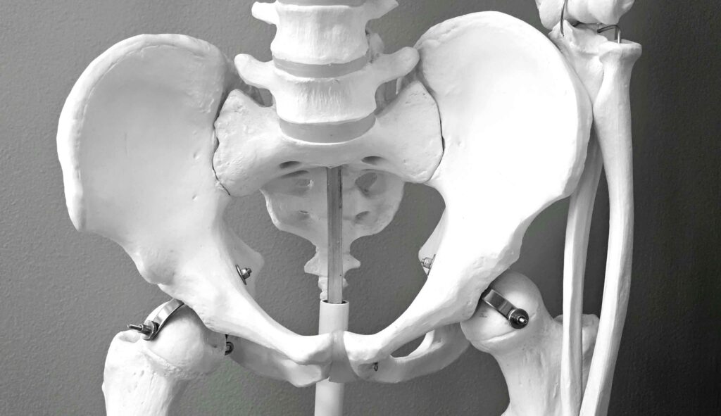
Anatomy of the Sacrum
Role of the Sacrum in the Human Body
To understand how sacral osteomyelitis develops and why it’s so critical, we first need to get familiar with the sacrum itself. The sacrum is a large, triangular bone located at the bottom of the spine, nestled between the two hip bones. It connects the spine to the pelvis and plays a key role in weight distribution during movement and while sitting or standing.
Structurally, the sacrum is composed of five fused vertebrae and acts as a keystone of the pelvic ring. It is surrounded by vital nerves, including the sacral plexus, which controls sensation and movement in the lower limbs. Because of its central position, an infection here can disrupt both musculoskeletal and neurological functions, leading to symptoms that may mimic other spinal or pelvic disorders.
Moreover, the sacrum’s rich vascular supply and porous nature make it susceptible to bacterial seeding—particularly from bloodborne infections or local spread. Once pathogens infiltrate the bone marrow, they can cause inflammation, pus accumulation, and destruction of bone tissue.
Why the Sacrum is Vulnerable to Infection
There are several reasons the sacrum is particularly vulnerable to osteomyelitis:
- Blood Supply: The sacrum receives blood from the median and lateral sacral arteries, which means bacteria from systemic infections can easily lodge there.
- Pressure Ulcers: In bedridden or immobile patients, the sacrum is a common site for pressure sores, which can become infected and spread to the underlying bone.
- Surgical Interventions: Procedures involving the spine or pelvis can introduce pathogens, especially if sterile protocols are breached.
- Proximity to GI and Urinary Tracts: Infections from the gastrointestinal or genitourinary tracts can potentially spread to the sacral area.
- Foreign Bodies and Implants: Devices like spinal stimulators or sacral screws can serve as a medium for bacterial colonization.
Understanding the sacrum’s anatomy and its vulnerabilities helps clinicians make a more accurate diagnosis and determine the best course of treatment.
Causes of Sacral Osteomyelitis
Hematogenous Spread
Hematogenous osteomyelitis refers to an infection that spreads through the bloodstream. In this scenario, bacteria like Staphylococcus aureus, Streptococcus, or even gram-negative organisms travel from a distant infection site—such as a urinary tract infection, skin abscess, or dental infection—and find their way to the sacrum. Once there, they settle in the bone’s vascular-rich marrow and begin to multiply, triggering an inflammatory response.
This is the most common route in children but also occurs in adults, especially those who are immunocompromised. Individuals with sepsis, IV drug use, or those undergoing hemodialysis are particularly at risk. The danger lies in the fact that the initial symptoms are vague—often just back pain or malaise—leading to a delay in diagnosis.
Left unchecked, hematogenous osteomyelitis can evolve rapidly, causing necrosis, abscess formation, and even bone collapse. Imaging and blood cultures are vital for early detection and intervention.
Direct Inoculation and Trauma
Another pathway through which infection can enter the sacrum is direct inoculation. This happens when there is an open wound or trauma involving the sacral area. Examples include:
- Open fractures
- Surgical incisions
- Penetrating injuries
- Spinal injections
Any breach in the skin or tissue barrier increases the risk of infection reaching the bone. Surgical procedures—particularly those involving spinal fusions or sacral hardware—are common culprits if aseptic techniques are not meticulously followed.
This type of osteomyelitis often presents more acutely, with redness, warmth, localized pain, and sometimes pus or fluid discharge from the wound. Prompt debridement and antibiotics are necessary to prevent the infection from spreading deeper.
Post-surgical and Pressure Ulcer-Related Infections
In patients who are bedridden, wheelchair-bound, or have undergone recent surgeries, the sacrum is a hotspot for pressure sores. When these ulcers become infected and aren’t properly managed, bacteria can infiltrate the underlying bone tissue.
Sacral pressure ulcers are notoriously difficult to treat once osteomyelitis sets in because the infection often goes undetected until it becomes chronic. These cases typically require aggressive wound care, imaging, bone biopsy, and prolonged antibiotics. In extreme situations, surgical removal of infected bone may be necessary.
This route of infection is particularly common in elderly individuals, spinal cord injury patients, and those in long-term care facilities. Prevention through mobility support, pressure-relieving surfaces, and skin care is key.
Common Risk Factors
Who is at Risk?
Understanding the risk factors associated with sacral osteomyelitis can help in early identification and prevention. While anyone can potentially develop it, certain populations are at a higher risk:
- Elderly Individuals: With weaker immune responses and a higher likelihood of comorbid conditions.
- Diabetics: Especially those with poor glycemic control, are highly susceptible to infections.
- Bedridden Patients: Prolonged immobility can lead to pressure ulcers that may become infected.
- IV Drug Users: Due to repeated bloodstream infections.
- Post-surgical Patients: Especially those with sacral instrumentation or spine surgeries.
- Cancer Patients: Particularly those on chemotherapy or immunosuppressive drugs.
Lifestyle and Health Conditions That Contribute
Aside from clinical vulnerabilities, lifestyle and health conditions also contribute significantly:
- Smoking: Reduces blood flow and impairs immune function.
- Obesity: Increases pressure on the sacrum and hinders mobility.
- Malnutrition: A weak immune system means a greater chance of infection.
- Alcoholism: Linked with liver disease and compromised immunity.
- Chronic Kidney Disease: Patients on dialysis have an increased risk of bloodborne infections.
A comprehensive evaluation of patient history and lifestyle is crucial in identifying those at risk and implementing preventive measures.
Symptoms and Early Warning Signs
Localized Pain and Swelling
One of the earliest and most persistent symptoms of sacral osteomyelitis is localized pain in the lower back or buttock area. It may start as a dull ache and gradually intensify. Patients often describe it as deep-seated or gnawing pain that doesn’t go away with rest or typical painkillers.
Swelling, redness, and warmth over the sacrum may also appear, particularly in cases of trauma or post-surgical infection. Sitting or lying down can exacerbate the discomfort, making daily tasks difficult. In some cases, the pain radiates to the thighs or legs, mimicking sciatica.
Mobility becomes limited, and patients might avoid physical activity, leading to further complications such as muscle atrophy and pressure ulcers.
Systemic Symptoms
While localized pain is a common and early sign, systemic symptoms are what often push people to seek medical attention. These include:
- Fever and Chills: Often a sign that the infection has spread beyond the localized area.
- Fatigue: Chronic infections like osteomyelitis can sap energy, making people feel unusually tired and weak.
- Night Sweats: A classic sign of a deep-seated infection.
- Loss of Appetite and Weight Loss: When your body is busy fighting an internal infection, appetite often takes a hit.
- Neurological Deficits: Numbness, tingling, or weakness in the lower limbs may occur if the infection compresses nearby nerves.
The problem is that these symptoms are often vague or resemble other conditions like disc herniation, sciatica, or even metastatic cancer. That’s why doctors need to maintain a high index of suspicion, especially in at-risk populations.
Blood tests may show elevated white blood cell counts, increased erythrocyte sedimentation rate (ESR), and high C-reactive protein (CRP) levels—all markers of inflammation and infection. However, imaging is usually necessary to confirm the diagnosis and determine the extent of bone involvement.
Diagnosis of Sacral Osteomyelitis
Physical Examination and Clinical History
Diagnosing sacral osteomyelitis requires a mix of detective work and clinical expertise. It starts with a detailed medical history, including recent infections, surgeries, injuries, and chronic conditions like diabetes or cancer. Doctors also assess for lifestyle risks such as prolonged immobility, IV drug use, or recent hospitalizations.
During the physical exam, they check for:
- Tenderness over the sacrum
- Swelling or redness in the lower back or buttocks
- Any visible signs of skin infection or pressure sores
- Signs of systemic infection like fever or rapid heart rate
A rectal or pelvic exam may be necessary to evaluate tenderness or masses suggestive of abscesses near the sacral region. Given its deep location, however, physical signs can often be subtle or completely absent.
Laboratory Tests and Markers
While imaging is crucial, lab tests provide the first clues:
- CBC (Complete Blood Count): May show elevated white blood cells.
- ESR and CRP: These inflammatory markers are often significantly raised in osteomyelitis.
- Blood Cultures: Help identify the organism causing the infection. Positive cultures can guide targeted antibiotic therapy.
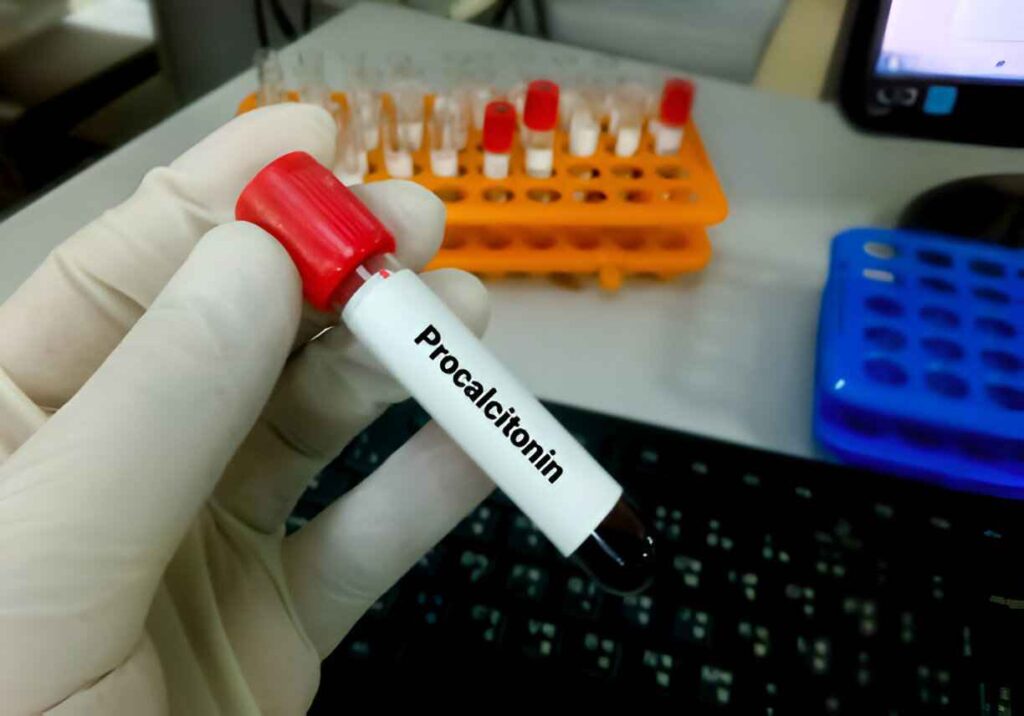
- Procalcitonin: A newer biomarker that may help distinguish bacterial infections from other inflammatory conditions.
A definitive diagnosis often requires a biopsy of the infected bone, particularly when blood cultures are negative. This can be done via CT-guided needle aspiration or during surgical debridement.
Imaging Techniques for Accurate Diagnosis
Imaging plays a central role in confirming sacral osteomyelitis:
- X-Rays: Often the first imaging test done, but early osteomyelitis may not show changes for up to 2 weeks.
- MRI (Magnetic Resonance Imaging): The gold standard for detecting osteomyelitis. It provides detailed images of bone marrow and soft tissues, and can show inflammation, abscesses, and necrosis.
- CT Scan: Useful for evaluating bone destruction and guiding biopsies.
- Bone Scan (Scintigraphy): Helps in detecting multifocal infections but lacks specificity.
- PET Scan: Highly sensitive for chronic infections and recurrence, particularly when MRI is inconclusive.
MRI findings typically show bone marrow edema, disc space narrowing, and soft tissue swelling. Gadolinium-enhanced scans can further delineate abscesses and infected tissue.
Treatment Options for Sacral Osteomyelitis
Antibiotic Therapy
The cornerstone of treatment for sacral osteomyelitis is antibiotics—often initiated even before culture results come back. Empiric therapy usually covers both gram-positive and gram-negative organisms. Once the offending pathogen is identified, treatment is tailored accordingly.
- Duration: Treatment typically lasts for 6–12 weeks.
- Route: Initially intravenous (IV), then may be switched to oral antibiotics if the patient shows improvement.
- Common Antibiotics Used:
- Vancomycin (for MRSA)
- Ceftriaxone
- Linezolid
- Ciprofloxacin
- Clindamycin
Patient response is monitored through clinical symptoms, repeat lab markers (CRP, ESR), and sometimes follow-up imaging.
It’s crucial not to stop treatment prematurely. Even if symptoms improve, stopping antibiotics early can lead to relapse or chronic osteomyelitis.
Surgical Interventions
Surgery is considered when:
- There is an abscess that needs drainage.
- The infected bone has died and needs removal (debridement).
- There’s spinal instability or risk of compression on spinal nerves.
- Antibiotics alone are insufficient after several weeks.
Surgical options may include:
- Debridement: Removal of infected tissue and necrotic bone.
- Abscess Drainage: Especially important when the infection involves surrounding soft tissues.
- Spinal Stabilization: In cases of structural compromise due to bone loss.
- Flap Coverage: For patients with pressure sores, plastic surgeons may perform soft tissue reconstructions using muscle or skin flaps.
Surgical management is often followed by prolonged antibiotic therapy and regular follow-up to prevent recurrence.
Recovery and Rehabilitation
Post-Treatment Care and Monitoring
Recovering from sacral osteomyelitis isn’t just about finishing a course of antibiotics or healing from surgery. It requires a well-structured post-treatment plan to prevent recurrence and rebuild strength. After initial treatment, patients are monitored closely to ensure the infection is completely eradicated.
Doctors will schedule regular follow-up visits and may recommend repeat imaging studies like MRI or CT scans to confirm healing of the bone and surrounding tissues. Lab tests—especially CRP and ESR—are used as indicators of inflammation and ongoing infection.
It’s also important to address any underlying conditions that may have contributed to the infection in the first place, such as uncontrolled diabetes or compromised immunity. Blood sugar control, immune-boosting diets, and treatment of chronic wounds or ulcers become vital parts of the recovery process.
Patients should also be educated about the warning signs of recurrence—return of pain, swelling, redness, or fever—and encouraged to seek prompt medical attention if symptoms return.
Physical Therapy and Mobility Support
Immobility is both a cause and a consequence of sacral osteomyelitis. That’s why rehabilitation is essential. Physical therapy can help patients:
- Regain mobility and strength
- Prevent pressure sores from reoccurring
- Improve posture and balance
- Minimize muscle atrophy due to prolonged inactivity
Depending on the severity of the case, therapy may include gentle stretching, assisted walking, or low-impact exercises. Occupational therapists may also work with patients to adjust their living environments—adding supportive cushions, adjusting bed positions, and teaching safe movement techniques.
In some cases, long-term mobility aids such as walkers, wheelchairs, or orthotic supports may be necessary, especially for elderly patients or those with concurrent spinal conditions.
Complications of Untreated Sacral Osteomyelitis
Chronic Infection and Abscess Formation
If sacral osteomyelitis isn’t diagnosed or treated early, it can become chronic. This means the infection lingers for months or years, creating ongoing inflammation and intermittent flare-ups. Chronic osteomyelitis often results in the formation of:
- Sequestrum: Dead bone tissue that becomes a reservoir for bacteria.
- Involucrum: New bone growth around the dead tissue, which can trap infection.
- Abscesses: Pockets of pus that may press on nerves and surrounding organs.
These conditions complicate treatment and often require surgical removal of the infected or dead tissue. Long-term antibiotics are also typically required, sometimes for life in non-operable cases.
Chronic infections weaken the immune system and can severely affect the patient’s quality of life, causing continuous pain, fatigue, and mobility issues.
Sepsis and Systemic Spread
One of the most life-threatening complications of untreated or poorly managed sacral osteomyelitis is sepsis. This occurs when the infection spreads from the bone to the bloodstream, causing a systemic inflammatory response.
Symptoms of sepsis include:
- High or low body temperature
- Rapid heartbeat
- Confusion or disorientation
- Shortness of breath
- Extremely low blood pressure (septic shock)
Sepsis requires immediate hospitalization and intensive care. Without prompt treatment, it can lead to multiple organ failure and death. This is why early diagnosis and proper management of sacral osteomyelitis are absolutely critical.
Preventing Sacral Osteomyelitis
Lifestyle Changes and Preventive Measures
Prevention is always better than cure—especially when it comes to a serious condition like sacral osteomyelitis. Some practical lifestyle changes and preventive measures include:
- Good Hygiene: Especially around wounds, pressure sores, or post-surgical sites.
- Frequent Position Changes: For bedbound patients, turning every 2 hours can help prevent pressure ulcers.
- Proper Nutrition: High-protein diets help with wound healing and immunity.
- Managing Chronic Illnesses: Controlling diabetes, kidney disease, and other health conditions reduces risk.
- Quit Smoking: Smoking impairs blood flow, which slows healing and increases infection risk.
Caregivers and healthcare professionals should be trained to identify early warning signs of pressure sores or skin infections—especially in high-risk populations.
Importance of Early Diagnosis and Intervention
Early detection is the key to preventing complications. Healthcare providers must stay vigilant, especially when dealing with high-risk groups. Patients experiencing unexplained back pain, especially if accompanied by fever or skin infections, should undergo a thorough evaluation.
Diagnostic imaging and lab tests should not be delayed. Starting antibiotics early—before the infection damages the bone irreversibly—can save lives and reduce the need for invasive surgeries.
Living with Sacral Osteomyelitis: Patient Perspectives
Coping Mechanisms and Support Systems
Living with sacral osteomyelitis is more than a medical journey—it’s an emotional and psychological challenge. Chronic pain, restricted mobility, and long recovery times can take a toll on mental health. That’s why coping mechanisms and support systems are essential.
- Support Groups: Joining a patient community or online forum can provide emotional support and valuable information.
- Counseling and Therapy: Professional guidance can help manage stress, depression, or anxiety related to long-term illness.
- Family and Caregiver Support: Having a strong network of caregivers and family members can make recovery smoother and less isolating.
Patients are also encouraged to set small, realistic recovery goals, track their progress, and celebrate small victories to maintain a positive outlook.
Success Stories and Long-Term Outcomes
Not every case of sacral osteomyelitis ends in surgery or permanent disability. Many patients recover fully, return to normal life, and resume their activities with no long-term complications. The key factors in these successful outcomes include:
- Early diagnosis
- Adherence to treatment
- Access to quality care
- Comprehensive rehabilitation
Each patient’s journey is unique, but hope and healing are entirely possible with the right approach and medical guidance.
Conclusion
Sacral osteomyelitis is a serious but treatable condition that demands attention, awareness, and timely intervention. While it may be rare, its consequences can be devastating if ignored or misdiagnosed. Understanding its causes, recognizing the early warning signs, and implementing a strong treatment and recovery plan can make all the difference.
Healthcare providers need to stay vigilant, especially for high-risk individuals, while patients should feel empowered to advocate for their health. With advancements in imaging, antibiotics, and surgical techniques, outcomes are improving. But nothing beats early detection and prevention.
Stay informed. Stay proactive. And don’t wait if something feels wrong—your health is too important to ignore.
FAQs
1. Is sacral osteomyelitis life-threatening?
Yes, it can be if not treated in time. The infection can spread to other parts of the body, leading to sepsis—a potentially fatal condition.
2. Can sacral osteomyelitis heal without surgery?
In many cases, yes. If caught early, antibiotic therapy alone may be sufficient. Surgery is generally reserved for more severe or chronic cases.
3. How long does it take to recover from sacral osteomyelitis?
Recovery time varies, but typically it ranges from 6 weeks to several months, depending on the severity and treatment approach.
4. What are the long-term effects of sacral osteomyelitis?
If treated effectively, many people recover fully. However, chronic infections can lead to long-term pain, reduced mobility, or the need for lifelong antibiotic therapy.
5. Who is most at risk of developing sacral osteomyelitis?
People with diabetes, weakened immune systems, those who are bedridden, or have undergone spinal surgeries are at the highest risk.