Atlantoaxial subluxation refers to the instability or partial dislocation between the first cervical vertebra (atlas) and the second (axis). This condition can cause neck pain, restricted movement, and in severe cases, compression of the spinal cord, which could lead to neurological symptoms. It might sound rare, but it’s more common among people with rheumatoid arthritis or children with Down syndrome.
Anatomy of the Atlantoaxial Joint
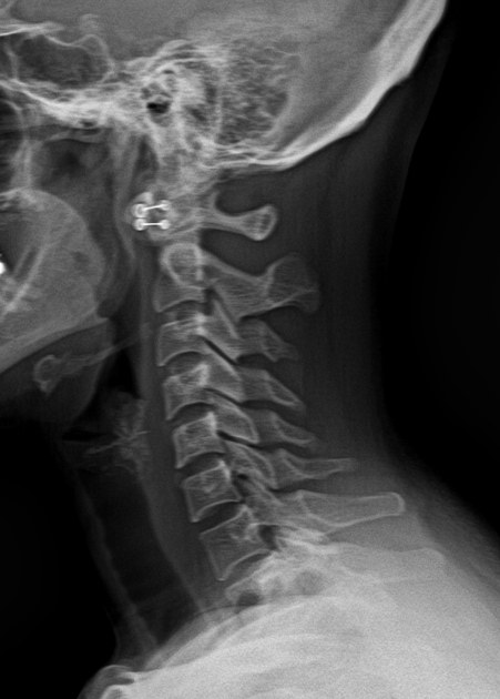
Details you can see in: https://jointshealth.site/cervical-disc-herniation/
To understand AAS, you need to get familiar with the unique anatomy of the atlantoaxial joint. The atlas (C1) and axis (C2) aren’t like other vertebrae in your spine. The atlas supports the skull and rotates around the dens, a bony projection from the axis. This rotation allows you to shake your head “no.”
The joint is held in place by several ligaments, especially the transverse ligament, which wraps around the dens like a seatbelt. If it’s damaged or becomes lax, the stability between C1 and C2 is compromised, which can lead to subluxation.
The atlas doesn’t have a vertebral body like the other vertebrae. Instead, it’s a ring that rotates around the dens. The entire setup is designed for movement, and almost 50% of your neck rotation happens at this joint.
Causes of Atlantoaxial Subluxation
Traumatic Causes
Accidents, especially those involving high-velocity impacts like car crashes or sports injuries, can cause direct trauma to the neck. The force can disrupt the ligaments holding C1 and C2 together, leading to immediate subluxation.
Inflammatory and Autoimmune Conditions
People with rheumatoid arthritis (RA) are especially prone to AAS. Inflammation wears down the ligaments and bones, particularly the transverse ligament. As these structures weaken, the atlantoaxial joint becomes unstable.
Congenital and Genetic Conditions
Children born with Down syndrome often have ligamentous laxity, meaning their ligaments are more stretchable and less capable of holding joints in place. This makes them more susceptible to AAS even without trauma. Other congenital anomalies like os odontoideum (a condition where the dens is detached) can also lead to instability.
Infectious or Post-surgical Causes
Infections like tuberculosis or surgical procedures around the cervical spine can weaken the structural integrity of the atlantoaxial joint. In rare cases, this can cause delayed onset AAS.
Understanding the root cause is vital because it dictates the treatment strategy, whether you need surgery or just conservative care.
Risk Factors and Populations at Risk
Certain groups are more vulnerable due to their medical history, genetics, or age.
Pediatric vs. Adult Onset
In children, especially those with genetic syndromes, the ligaments are more elastic. This means they’re naturally more prone to joint instability. Atlantoaxial subluxation in kids often stems from congenital or developmental issues. On the flip side, adults usually develop AAS due to trauma or inflammatory diseases.
Rheumatologic Conditions
Studies show that up to 50% of patients with rheumatoid arthritis may develop some form of cervical spine instability over time. The chronic inflammation erodes ligaments and bones, making the joint wobbly and dangerous.
Genetic Syndromes
Conditions like Down syndrome and Morquio syndrome come with built-in risks. These syndromes often feature ligamentous laxity or bony anomalies, setting the stage for potential AAS. For this reason, routine screening for neck instability is often recommended in kids with these conditions, especially before surgeries or intense physical activity.
Signs and Symptoms
You might think neck pain is the most obvious symptom of AAS, and you’d be right. But that’s just the tip of the iceberg.
Neck Pain and Limited Movement
This is usually the first red flag. Patients often complain of stiffness, soreness, or a dull ache at the base of the skull. Some people describe a feeling of instability, like their head isn’t sitting right on their neck.
Neurological Symptoms
As the subluxation worsens, it can compress the spinal cord. This is where things get serious. Patients may experience:
- Numbness or tingling in the arms or legs
- Loss of coordination
- Weakness
- Difficulty walking
Signs of Cervical Myelopathy
In advanced cases, the spinal cord itself is compressed, leading to myelopathy, a condition that can affect your bladder, bowel, and even respiratory function. These signs include:
- Loss of fine motor skills
- Clumsiness in the hands
- Spasticity in the legs
The longer the spinal cord is compressed, the higher the risk of permanent damage. If you, or someone you know, is experiencing these signs, it’s time to see a doctor, fast.
Diagnosis of Atlantoaxial Subluxation
Diagnosing this isn’t just about snapping an X-ray and calling it a day. This condition requires a thorough approach that involves a combination of physical assessment and detailed imaging to confirm the extent of instability and its impact on the spinal cord.
Physical Examination
It usually starts with a clinical evaluation. A skilled clinician will check for:
- Neck tenderness
- Reduced range of motion
- Muscle weakness
- Neurological signs such as reflex abnormalities or coordination issues
They might also assess posture, gait, and balance, especially if spinal cord involvement is suspected. In children, signs like a head tilt or torticollis could also raise suspicions.
Imaging Techniques
Here’s where things get more precise. Dynamic cervical spine X-rays (taken in flexion and extension) are often the first step. These show how much movement occurs between the atlas and axis and can reveal abnormal gaps between bones, especially the atlantodental interval (ADI).
But for a more detailed look, doctors will order a CT scan or MRI:
- CT scans give a clear picture of the bony anatomy and are great for detecting fractures or congenital malformations.
- MRI is crucial when spinal cord compression is suspected. It helps visualize soft tissues, including ligaments and the spinal cord itself.
Neurological Testing
When symptoms suggest nerve involvement, an electromyography (EMG) or nerve conduction study might be performed to check nerve function.
Accurate diagnosis is the cornerstone of effective treatment. Early detection can prevent complications, especially in at-risk populations like children with Down syndrome or adults with autoimmune diseases.
Classification of Atlantoaxial Subluxation
Understanding the type and severity helps in choosing the right treatment path. Medical professionals use various classification systems to describe the condition more precisely.
Rotatory vs. Non-rotatory Subluxation
- Rotatory AAS: This is when the atlas (C1) rotates abnormally on the axis (C2). It often happens in children and may be caused by minor trauma or inflammation.
- Non-rotatory AAS: This form is more common in adults and typically results from ligament laxity or erosion from conditions like rheumatoid arthritis.
Fielding and Hawkins Classification
This system is commonly used for rotatory subluxation and breaks it down into four types:
- Type I: Simple rotatory subluxation without anterior displacement.
- Type II: Rotation with slight anterior displacement (3-5mm).
- Type III: Rotation with more significant anterior displacement (>5mm).
- Type IV: Rotation with posterior displacement, often more severe and rare.
Severity Grading Based on ADI
The atlantodental interval (ADI) is a key measure:
- Normal: Less than 3mm in adults; less than 5mm in children.
- Mild: Slight widening, often without neurological symptoms.
- Severe: ADI greater than 5mm, usually with signs of spinal cord compression.
These classifications aren’t just academic—they guide treatment. For example, mild cases may be managed conservatively, while severe or unstable ones often require surgery.
Complications of Untreated Subluxation
Let’s get one thing straight: untreated atlantoaxial subluxation is dangerous. What starts as neck stiffness or mild pain can snowball into catastrophic outcomes if the condition progresses unchecked.
Spinal Cord Compression
This is the most alarming complication. As the vertebrae shift, they can narrow the spinal canal and squeeze the spinal cord. This can lead to:
- Paralysis
- Loss of bladder or bowel control
- Difficulty breathing in extreme cases
Once nerve tissue is damaged, recovery becomes difficult or impossible, making timely treatment crucial.
Chronic Pain and Reduced Mobility
Even if the spinal cord isn’t compressed, the instability can cause ongoing pain, muscle spasms, and restricted motion. This affects quality of life and may lead to long-term dependence on painkillers or other interventions.
Neurological Deterioration
As nerve function declines, patients may experience progressive symptoms:
- Tingling or numbness in limbs
- Muscle weakness
- Balance and coordination issues
In children, untreated subluxation can interfere with development, cause behavioral changes due to chronic discomfort, and even lead to permanent deformities.
In short, this isn’t something you can ignore or “tough out.” Without proper care, the condition only worsens—often with irreversible consequences.
Non-Surgical Treatment Options
Not every case of AAS demands a scalpel. In fact, many mild to moderate cases respond well to non-surgical management, especially if diagnosed early.
Immobilization and Bracing
For patients with minimal subluxation and no neurological deficits, cervical collars or halo vests can be used to stabilize the neck and promote healing. This is particularly effective in kids with rotatory subluxation.
Physical Therapy
Under careful supervision, physical therapy can help:
- Strengthen neck muscles
- Improve posture
- Enhance stability
However, aggressive or unsupervised movements are a no-go—they can worsen the subluxation.
Medications
Anti-inflammatory drugs, like NSAIDs or corticosteroids, are often prescribed to reduce inflammation, especially in inflammatory types of AAS. In RA patients, disease-modifying antirheumatic drugs (DMARDs) or biologics may help control the underlying condition.
Manual Therapy
In rare, very mild cases, some manual therapy techniques may help realign the joint. But this is controversial and should only be done by highly trained professionals. Improper manipulation could cause more harm than good.
Non-surgical treatments offer relief without the risks of surgery, but they’re not for everyone. Regular monitoring is essential to ensure the condition doesn’t deteriorate under conservative management.
Surgical Treatment Options
When non-surgical routes don’t cut it, especially in severe or progressing cases, surgery becomes the best option. The goal is to stabilize the joint, relieve pressure on the spinal cord, and prevent further damage.
Indications for Surgery
You may need surgery if:
- There is significant spinal cord compression
- ADI exceeds 5mm
- Non-surgical treatments fail
- Symptoms are progressively worsening
Common Surgical Procedures
- Posterior Fusion (C1-C2 Fusion): The most common technique, where screws and rods are used to stabilize the vertebrae. Bone grafts help fuse the bones together over time.
- Decompression Surgery: If there’s pressure on the spinal cord, surgeons may remove part of the bone or disc to relieve it.
- Transoral Odontoid Resection: Rare but used when the dens is pressing on the spinal cord from the front. This procedure is complex and often followed by fusion.
Recovery and Risks
Post-surgery, patients typically wear a cervical collar and undergo physical therapy. Recovery time varies but often takes several months. As with any surgery, there are risks:
- Infection
- Nerve damage
- Hardware failure
That said, most patients see dramatic improvements in pain and neurological symptoms after surgery. With proper rehab and care, many return to a relatively normal life.
Atlantoaxial Subluxation in Children
In children, this condition presents unique challenges, both in diagnosis and treatment. Their growing bodies and developing anatomy make it critical to catch this condition early.
Children’s ligaments are naturally more flexible, and their bones are still forming. This flexibility increases their range of motion but also makes them more susceptible to joint instability. Kids with Down syndrome, in particular, are at high risk because of ligamentous laxity and potential congenital abnormalities in the cervical spine.
Other conditions like Grisel’s syndrome (a rare non-traumatic rotatory AAS linked to infections of the head or neck) can also lead to subluxation. Unlike adults, children may not express neck pain clearly but may show signs like:
- Persistent head tilt
- Reluctance to move the head or neck
- Irritability or crying when turning the head
Diagnosis Challenges
Imaging is essential but must be done carefully to minimize radiation exposure. Pediatric-specific techniques like dynamic X-rays with close monitoring are preferred. MRI is ideal if neurological symptoms are suspected, especially to assess soft tissue involvement.
Treatment Approaches
Conservative treatment works well in most pediatric cases:
- Cervical collars
- Traction therapy
- Physical therapy with gentle exercises
In more severe cases or if conservative methods fail, surgical stabilization may be needed. Children typically heal faster than adults and have a better long-term prognosis if treated promptly.
Preventive Screening
Due to the known risks, especially in children with Down syndrome, routine screening is often done around age 3-5, before they participate in activities like gymnastics or undergo surgery requiring general anesthesia. Early detection helps prevent serious complications later in life.
Prognosis and Long-term Outlook
The prognosis varies depending on the cause, severity, and the stage at which it’s caught.
When caught early, especially before neurological symptoms develop, the prognosis is generally excellent. Conservative treatments like bracing and physical therapy often resolve the issue without lasting damage. Even surgical outcomes are typically good, with many patients returning to normal activities within a few months.
If left untreated, AAS can lead to:
- Chronic pain
- Progressive neurological symptoms
- Spinal cord damage
In extreme cases, the compression can affect vital functions such as breathing and mobility, severely impacting quality of life.
Risk of Recurrence
Recurrence is rare if the underlying cause is addressed, but it’s not impossible. Patients with autoimmune disorders or congenital conditions require long-term monitoring. Follow-up imaging every few years may be necessary to ensure the spine remains stable.
Living with Atlantoaxial Subluxation
Living with AAS, even after successful treatment, requires adjustments. But with the right habits and awareness, you can still lead a full, active life.
- Avoid high-impact sports: Activities like football, wrestling, or trampoline jumping can be dangerous due to the risk of trauma.
- Use supportive devices: Special pillows for sleep, neck braces during flare-ups, and ergonomic chairs can help.
- Regular check-ups: Consistent follow-ups with a spine specialist or rheumatologist ensure that the condition remains under control.
Managing Chronic Symptoms
Some people may continue to experience residual symptoms like neck stiffness, mild pain, or fatigue. These can be managed with:
- Heat and cold therapy
- Gentle stretching and low-impact exercises like yoga or swimming
- Mindfulness and stress management techniques
Emotional and Mental Health
Chronic conditions often affect mental well-being. Dealing with pain, limited mobility, or the fear of worsening symptoms can lead to anxiety or depression. It’s important to seek emotional support:
- Talk therapy
- Support groups (especially helpful for parents of children with AAS)
- Family counseling
Being proactive about mental health can make a huge difference in overall quality of life.
Prevention Tips and Early Detection
You can’t always prevent AAS, but knowing the risks and taking the right precautions can dramatically reduce the odds of developing severe symptoms.
Monitor High-Risk Individuals
If you or your child have any of the following, regular monitoring is essential:
- Rheumatoid arthritis
- Down syndrome
- History of neck trauma
- Congenital cervical anomalies
Annual or biannual imaging may be recommended in some cases.
Protect the Neck
- Always wear seatbelts and use appropriate car seats for children.
- Wear helmets during activities like biking or skateboarding.
- Avoid sudden, jerky movements of the neck.
Infection Management
In children, upper respiratory infections like tonsillitis can trigger inflammatory responses that lead to subluxation (as in Grisel’s syndrome). Prompt treatment of infections can reduce this risk.
Educate and Advocate
If you or your child are in a high-risk group, make sure every doctor, physical therapist, and coach knows about the condition. Proper education ensures that everyone involved can take necessary precautions.
When to See a Doctor
Knowing when to get help can make all the difference in preventing serious complications. If you experience—or notice in a child—any of the following, don’t wait:
Red Flag Symptoms
- Persistent neck pain or stiffness
- Trouble turning the head
- Numbness or tingling in the arms or legs
- Loss of coordination or clumsiness
- Difficulty walking or balancing
- Sudden weakness in the limbs
Emergency Symptoms
- Trouble breathing
- Loss of bladder or bowel control
- Severe headache or dizziness
These could indicate spinal cord compression, a medical emergency requiring immediate attention.
Who to See
Start with a primary care provider, who may refer you to:
- Neurologist
- Orthopedic surgeon
- Rheumatologist (especially for autoimmune causes)
- Pediatric specialist (for children)
Getting a second opinion from a spine specialist is never a bad idea—especially when surgery is being considered.
Conclusion
Atlantoaxial subluxation, whether caused by trauma, genetics, or autoimmune disease, requires early recognition and treatment. With modern diagnostic tools, advanced surgical options, and a growing understanding of the condition, outcomes have improved.
From children born with genetic vulnerabilities to adults developing instability due to arthritis, awareness is your first line of defense. Know the signs, get regular check-ups if you’re at risk, and never ignore neck pain that doesn’t go away.